

Mal del ciprés, stages of decline
Photo Gallery
Site will be retired 9/1/2026
This site is no longer being developed and will be retired on September 1, 2026. Please contact us if you have any questions or would like to provide support to continue the project.
|
P. austrocedrae - Mal del ciprés, stages of decline 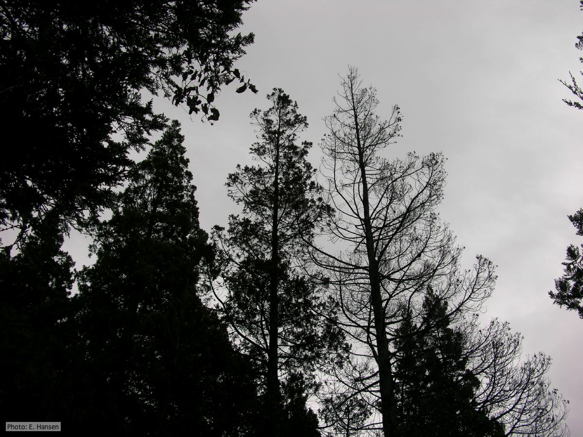 |
P. cinnamomi hyphal swellings 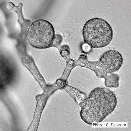 P. cinnamomi hyphal swellings (or thin walled chlamydospores) |
P. ramorum sporangium  Deciduous sporangium, photo from Q-bank, used with permission |
|
P. ramorum sporangia and chlamydospores 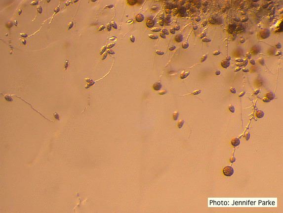 Sporangia and chlamydospores of P. ramorum |
P. arenaria hyphal swellings 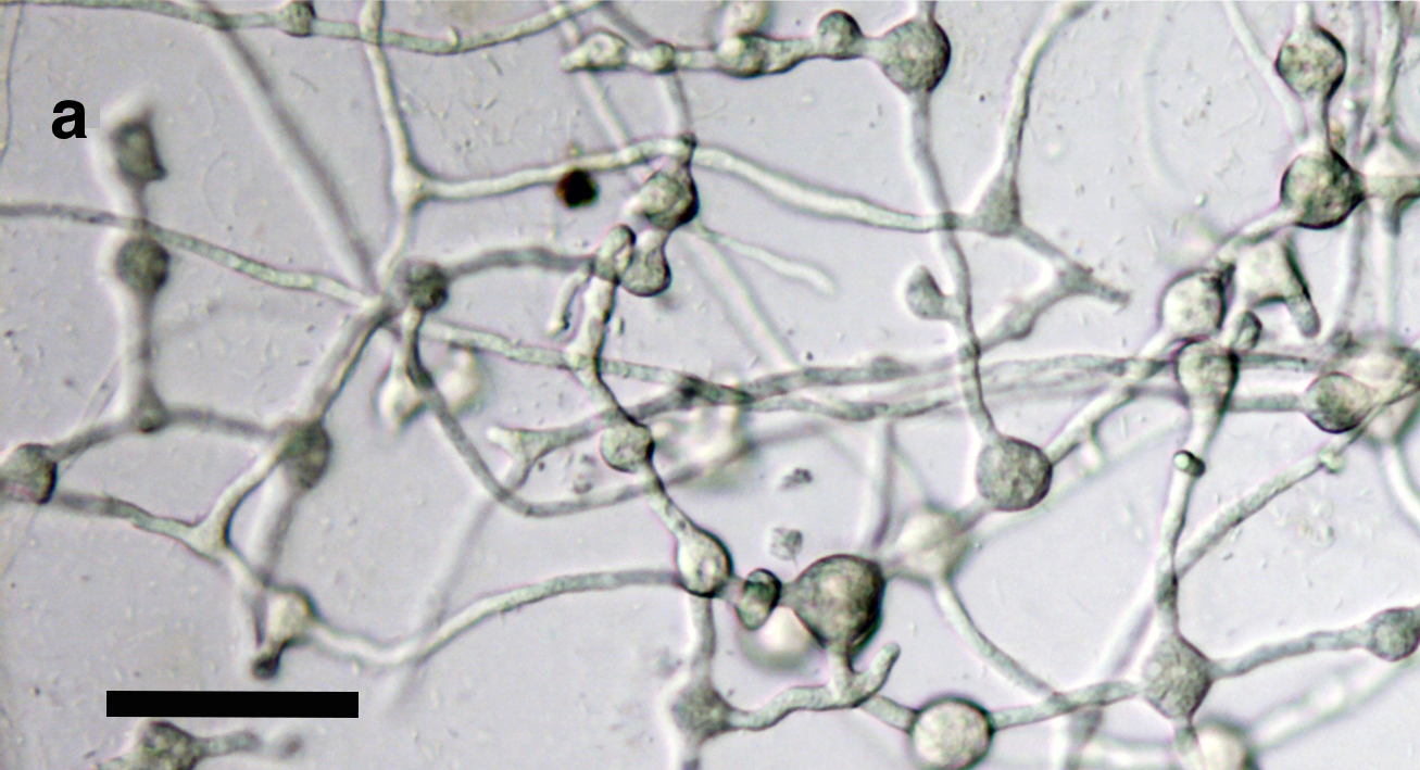 Catenulate, globose to subglobose hyphal swellings, some of them with radiating hyphae scale bar = 50 μm. |
P. cactorum sporangium  P. cactorum sporangium |
|
Boot wash to station to control spread of P. agathidicida 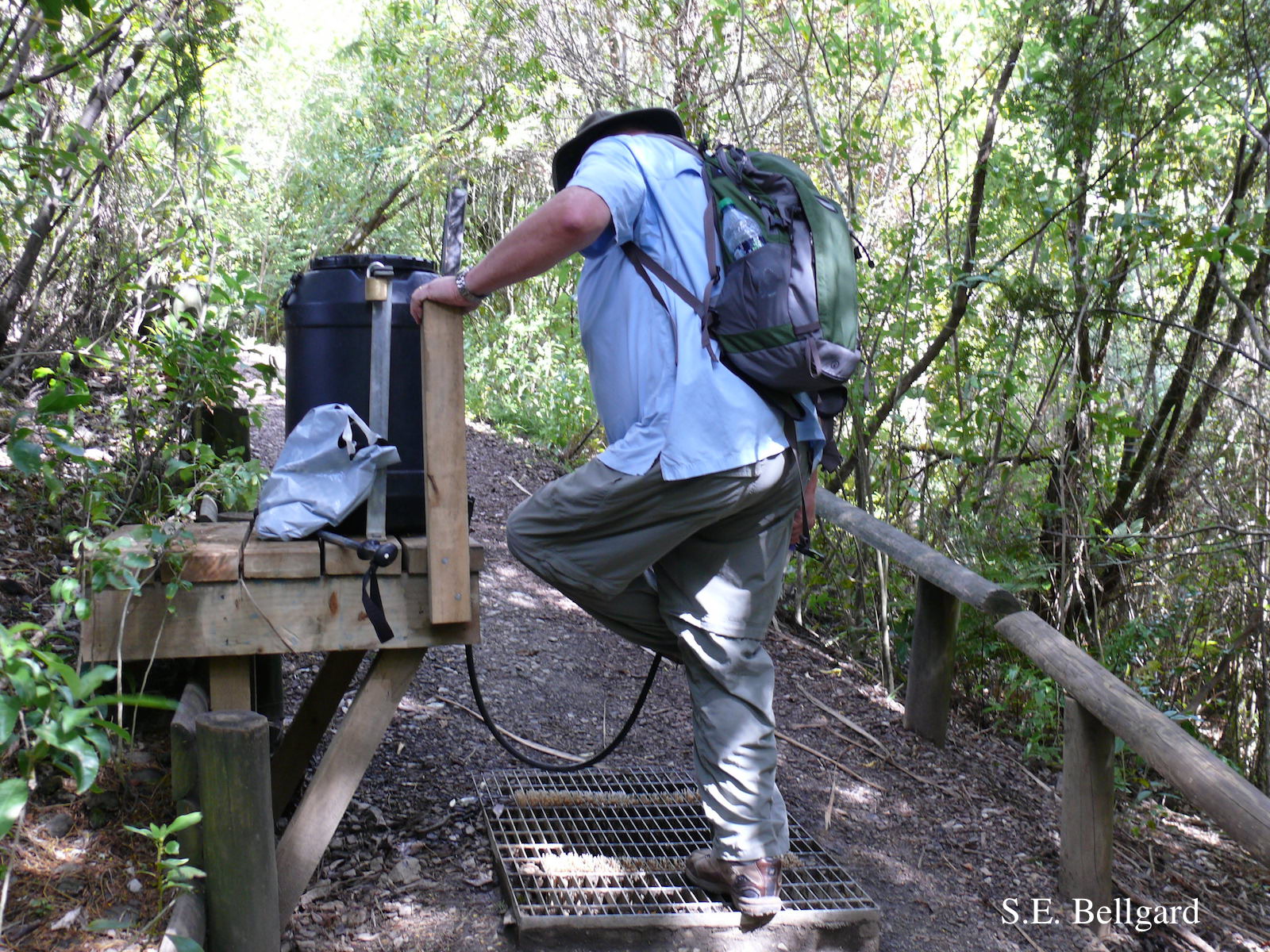 Use of hypochlorite solution applied through a “livestock drench-gun”, integrated with a soil grate to allow potentially contaminated soil to be collected |
P. chlamydospora sporangium 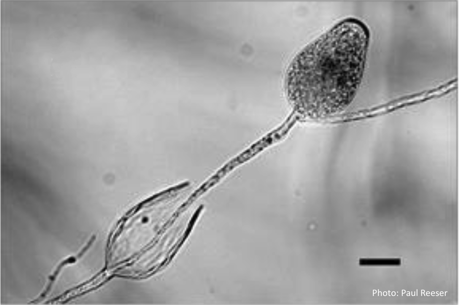 Phytophthora chlamydospora sporangia in water, showing subsporangial elongation. Bar is 20 µm. |
P. cambivora sporangium 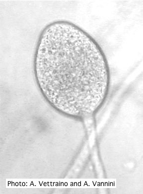 Ovoid non-papillate sporangia with well-rounded base and simple sporangiophore |
|
P. frigida sporangia 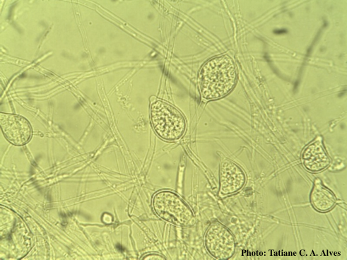 Noncaducous sporangia showing ovoid shape and papillate condition |
P. kernoviae leaf wilt 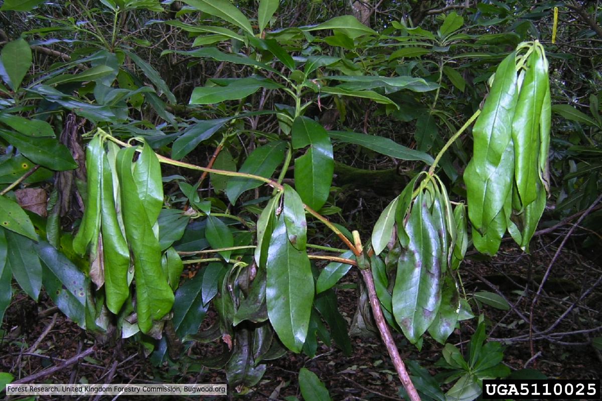 Wilted leaf of infected rhododendron |
P. pseudosyringae sporangia  Ovoid, semipapillate sporangia showing sympodial development of sporangiophore |