

Fagus sylvatica bole canker
Photo Gallery
Site will be retired 9/1/2026
This site is no longer being developed and will be retired on September 1, 2026. Please contact us if you have any questions or would like to provide support to continue the project.
|
P. cambivora bole canker 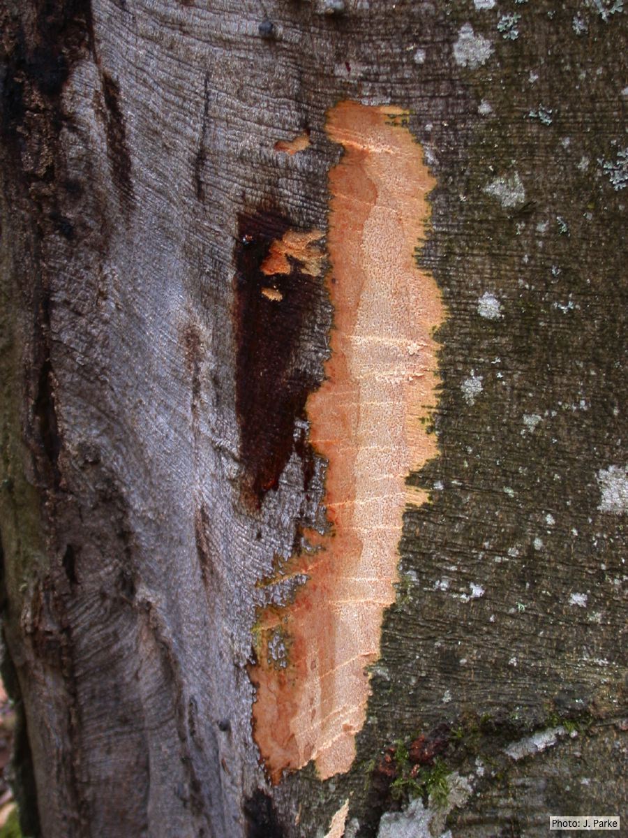 |
P. alni in alder forest, Germany, with T. Jung 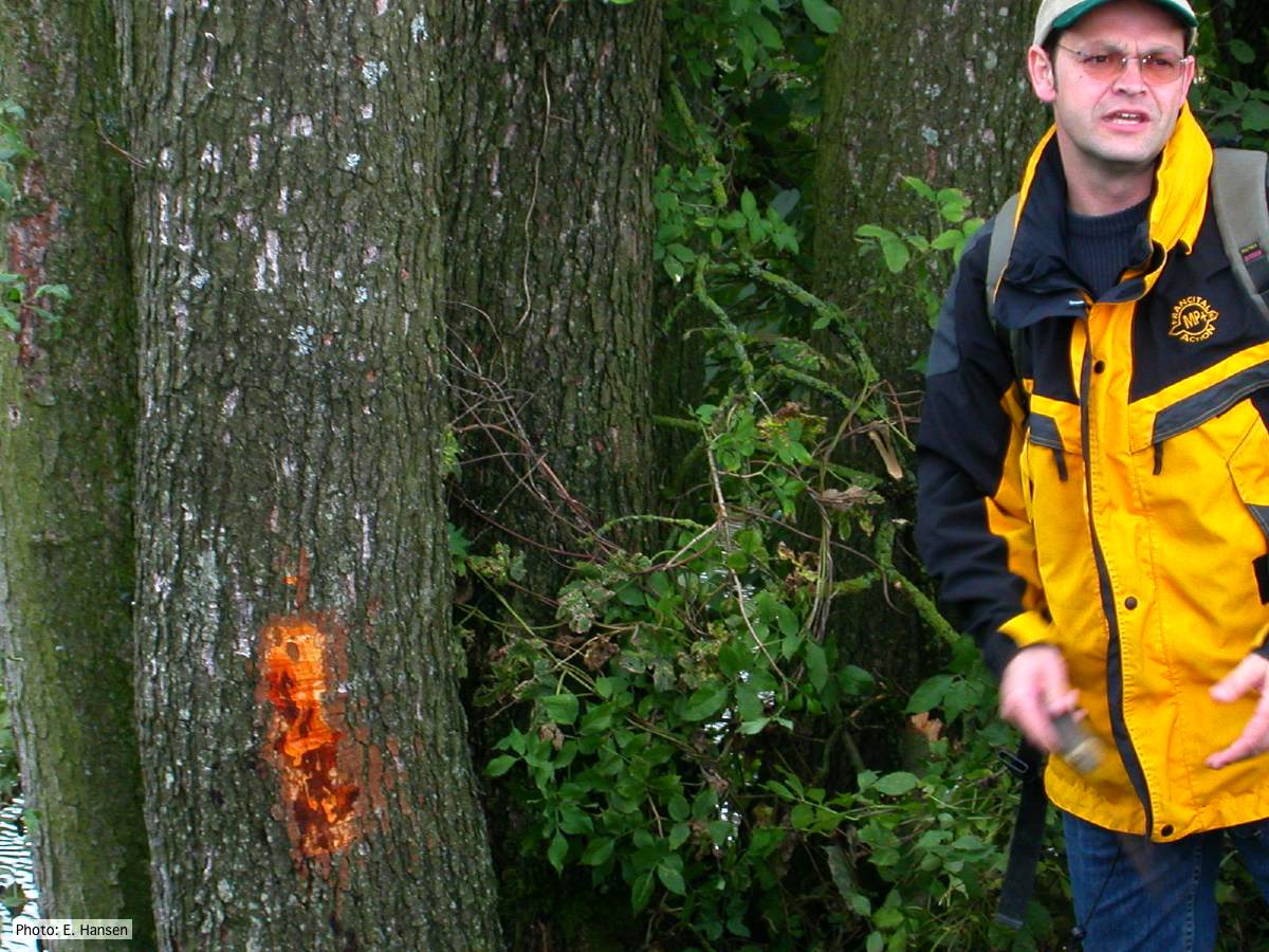 P. alni in alder forest, Germany, with T. Jung |
P. pseudosyringae sporangia  Ovoid, semipapillate sporangia showing sympodial development of sporangiophore |
|
P. kernoviae canker  Bole lesion on Fagus sylvatica |
P. megakarya oospore
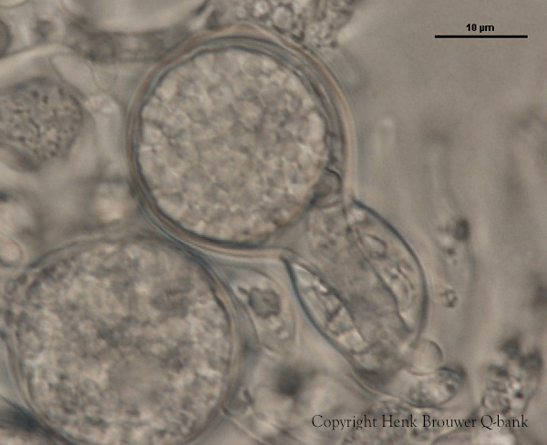
P. megakarya oogonia, oospore, and antheridium
|
P. pseudosyringae sporangium 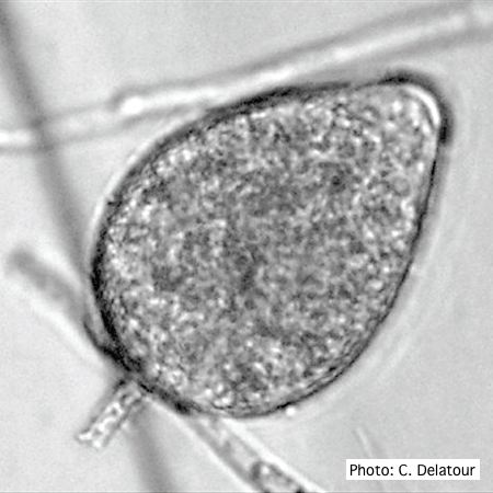 Ovoid, semipapillate sporangia showing medium length pedicel |
|
P. megakarya disease symptoms on Theobroma cacao fruit 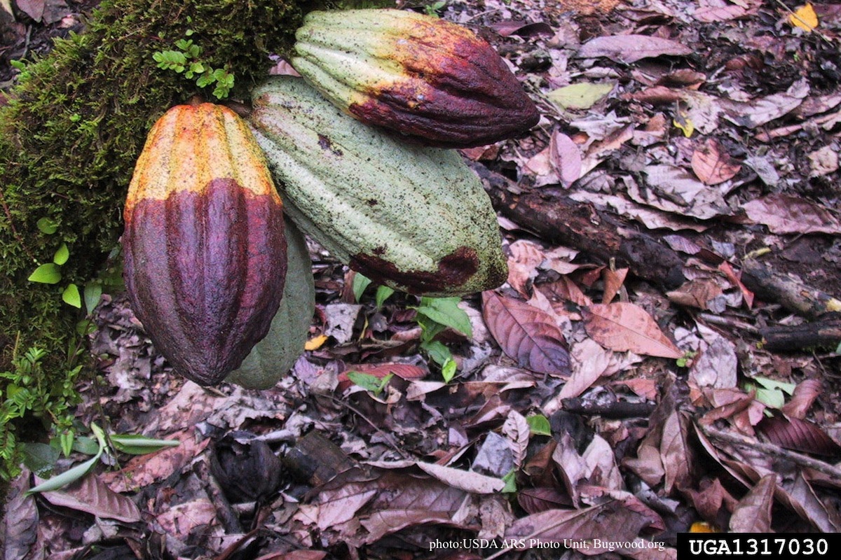 Disease symptoms on a cocoa pod |
P. cryptogea colony morpholgy on V8  Colony morphology on V8 at 14 days |
P. cinnamomi colony morphology on PDA  P. cinnamomi colony growth on PDA at 14 days |
|
P. cactorum colony morphology on PDA 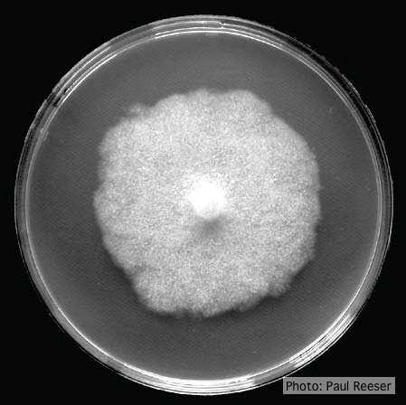 Colony morphology on PDA at 14 days |
P. cinnamomi hyphal swelling 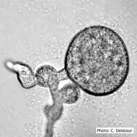 P. cinnamomi hyphal swelling (or thin walled chlamydospores) |
Comparative gametangial morphology of Phytophthora Clade 5 species  Comparative gametangial morphology of Phytophthora Clade 5 species, with SEM (top) and light microscopy (bottom). P. heveae has smooth walled oogonia with funnel-shaped, amphigynous antheridia. P. agathidicida has mildly stipulate oogonia with globose amphigynous antheridia. P.cocois has mildly bullate oogonia with reflexed amphigynous antheridia. P. castaneae has coarsely bullate oogonium with rugose protuberances and narrow amphigynous antheridia (Weir et al. 2015). |