

Coenocytic hyphae (from Duran et al. 2008). Scale bar = 20 μm.
Photo Gallery
Site will be retired 9/1/2026
This site is no longer being developed and will be retired on September 1, 2026. Please contact us if you have any questions or would like to provide support to continue the project.
|
P. pinifolia coenocytic hyphae 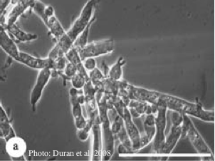 |
P. nemorosa oogonium 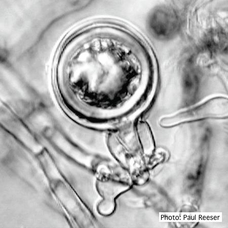 Oogonium with amphigynous antheridium |
P. siskiyouensis canker on Italian alder 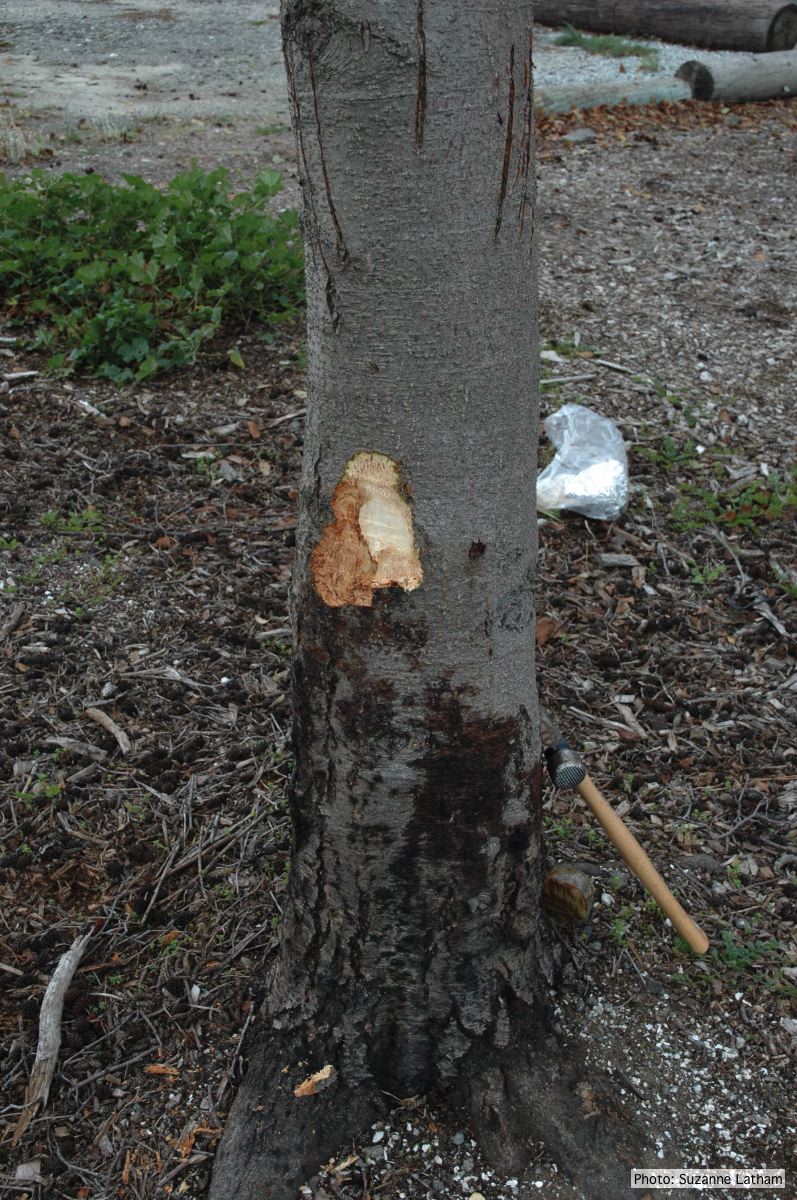 Bole lesions in the tissues under the bark of a bleeding canker: distinct margin between healthy and disease tissues |
|
P. pinifolia on Pinus radiata 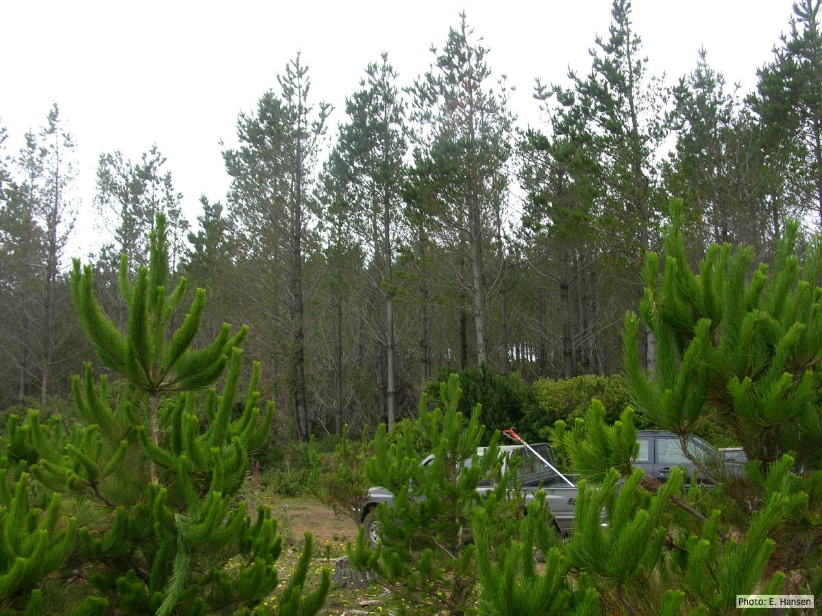 Pinus radiata stand, note Defoliation and regrowth |
P. chlamydospora chlamydospore 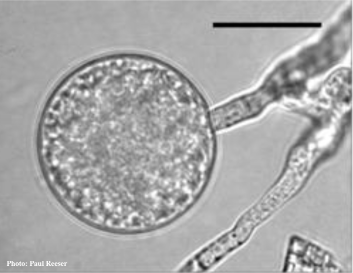 Phytophthora chlamydospora chlamydospore in agar. Bar is 20 µm. |
Growth of P. megakarya on PDA 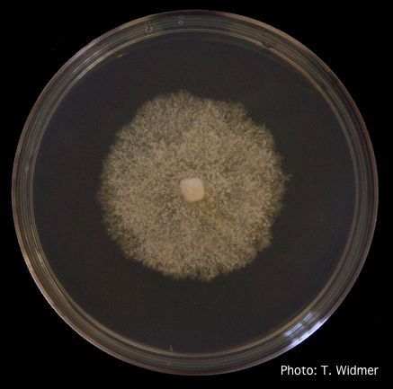 Growth of P. megakarya on potato dextrose agar |
|
P. agathidicida oospores in planta  Oospores in the roots of kauri seedlings inoculated with P. agathidicida. The root has been cleared with potassium hydroxide and bleached with peroxide before being stained with trypan blue (scale bar =100 µm). |
P. ramorum colony morphology on V8 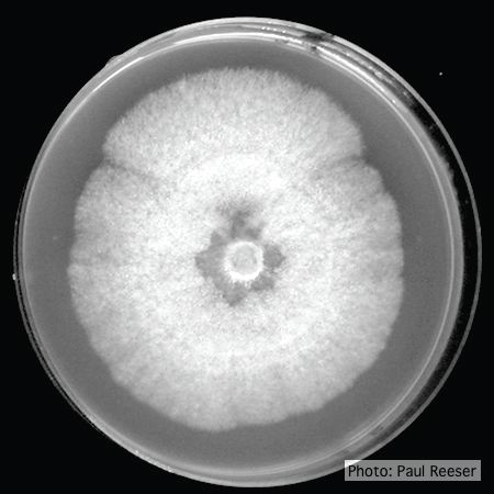 P. ramorum colony morphology on V8 |
P. siskiyouensis disease symptoms on Italian alder 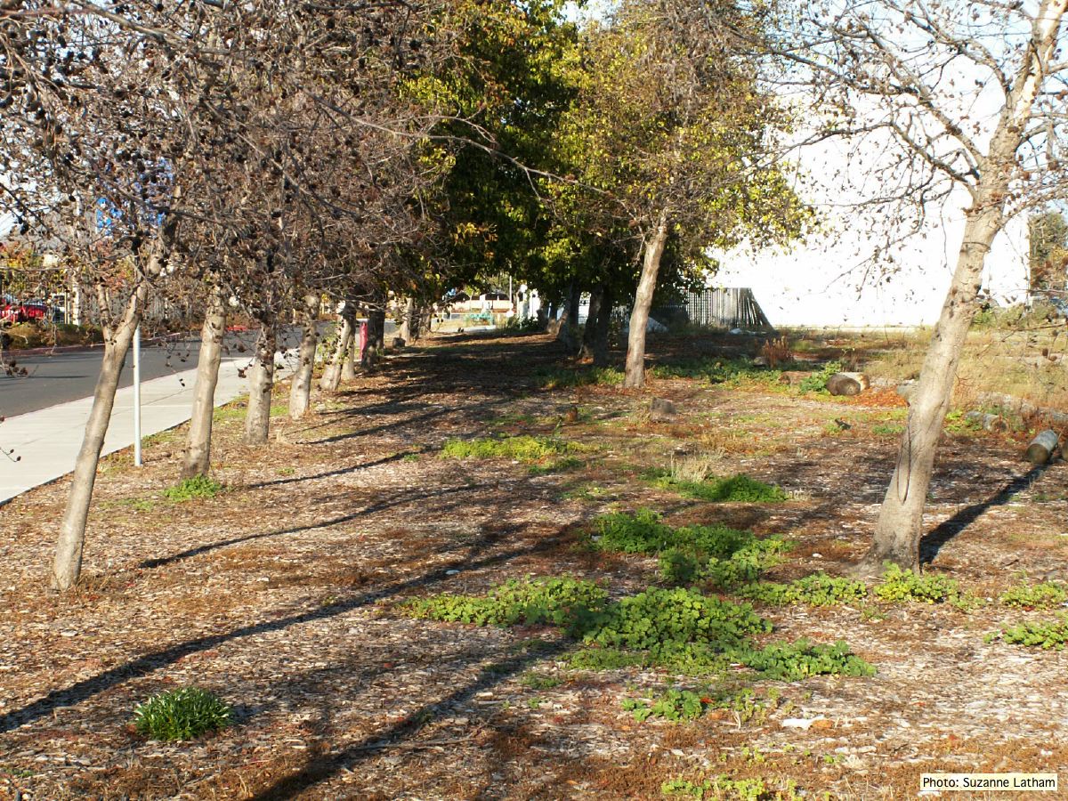 Grove of dying trees in a commercial landscape in Foster City, CA |
|
P. pluvialis oogonium and antheridium  Oogonium and oospore with amphigynous antheridium |
P. austrocedrae colony morphology on Tomato juice agar with B sitosterol 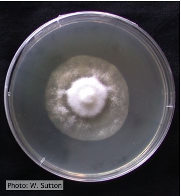 Colony morphology of P. austrocedrae at 16 ºC after 4 weeks on Tomato juice agar with B sitosterol |
Black pod disease of cacao caused by P. palmivora 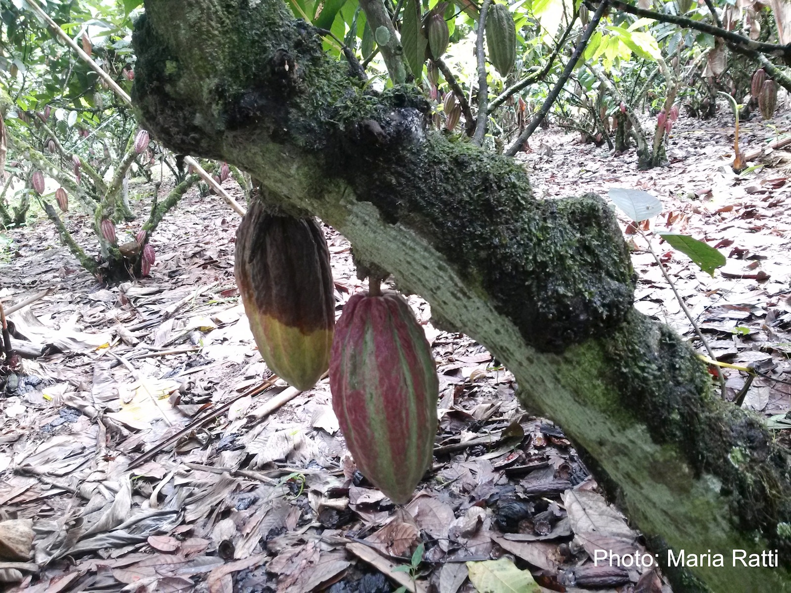 Black pod of cacao in Ecuador caused by P. palmivora (see lesioned fruit on left). |