

Non- papillate and caducous sporangia of Phytophthora pinifolia isolated from the infected P. radiata needles.
Photo Gallery
Site will be retired 9/1/2026
This site is no longer being developed and will be retired on September 1, 2026. Please contact us if you have any questions or would like to provide support to continue the project.
|
P. pinifolia sporangium 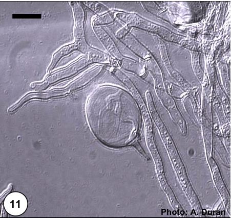 |
P. megasperma colony morphology on V8  Colony morphology on V8 at 7 days |
P. cinnamomi on Jarrah 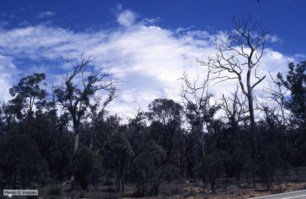 Dieback in Jarrah, Western Australia |
|
P. alni canker on gray alder 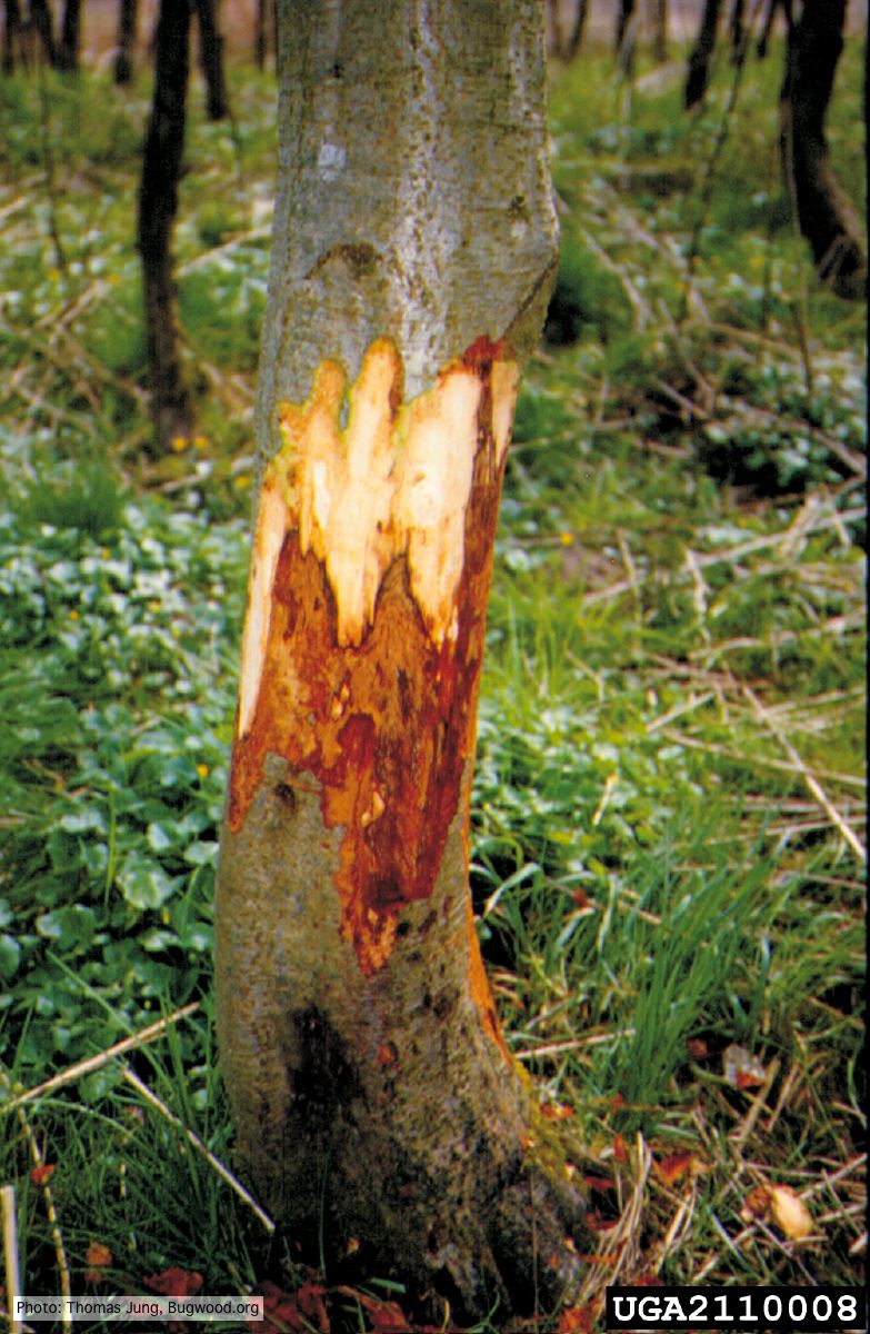 Grey alder (A. incana) with collar rot caused by P. alni |
Phytophthora cactorum disease symptoms on English walnut  Dieback of Juglans regia caused by Phytophthora cactorum |
P. boehmeriae sporangium 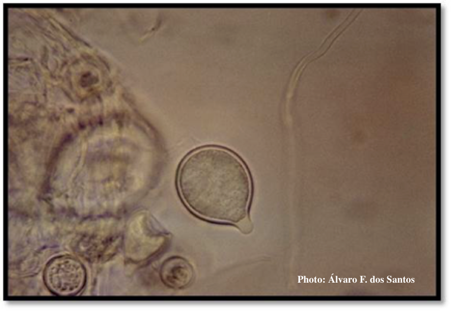 Sporangium showing ovoid and ovoid to spherical shape and papillate condition |
|
P. agathidicia sporangia 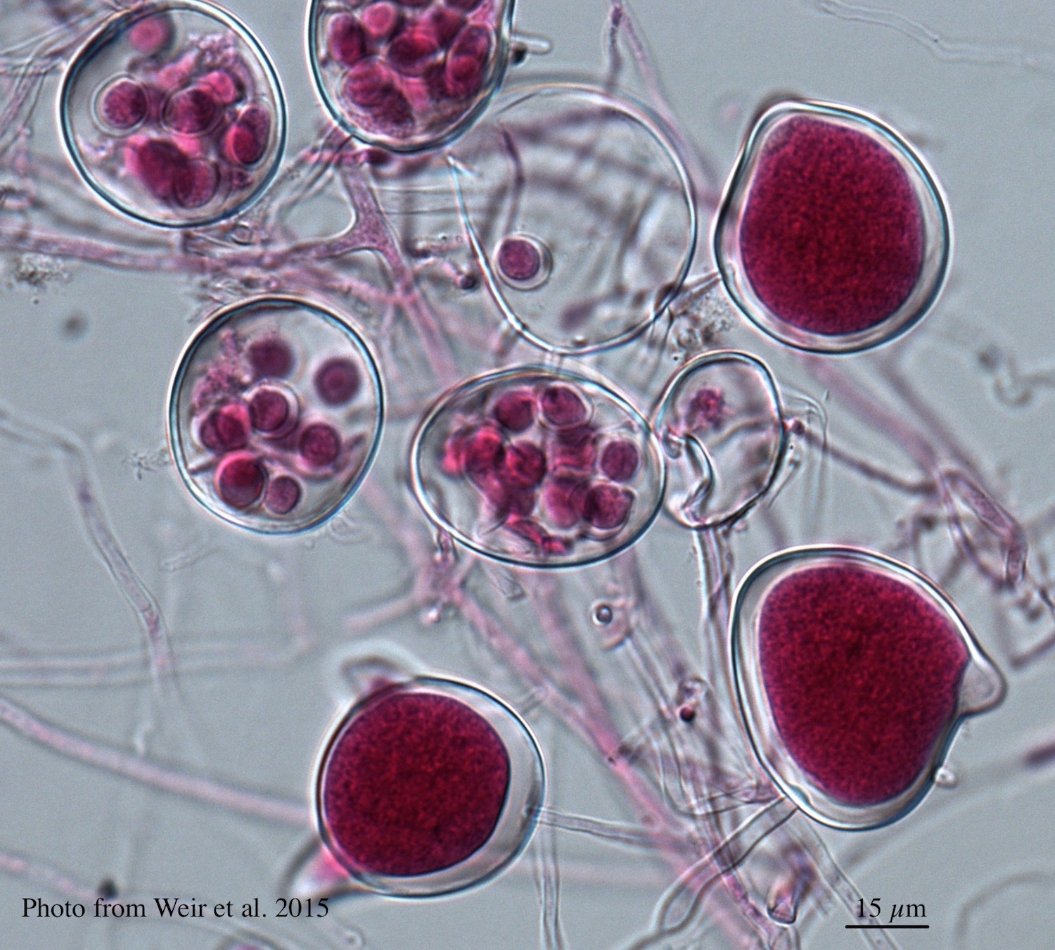 Differentiation of the cytoplasm within papillate sporangia into acid fuchsin stained zoospores |
P. katsurae disease symptoms  Infected chestnut (Castanea) with bleeding canker |
P. cryptogea colony morpholgy on PDA 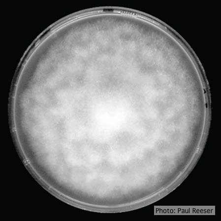 Colony morphology on PDA at 14 days |
|
Comparative gametangial morphology of Phytophthora Clade 5 species  Comparative gametangial morphology of Phytophthora Clade 5 species, with SEM (top) and light microscopy (bottom). P. heveae has smooth walled oogonia with funnel-shaped, amphigynous antheridia. P. agathidicida has mildly stipulate oogonia with globose amphigynous antheridia. P.cocois has mildly bullate oogonia with reflexed amphigynous antheridia. P. castaneae has coarsely bullate oogonium with rugose protuberances and narrow amphigynous antheridia (Weir et al. 2015). |
P. kernoviae colony morphology on CMA PARPH 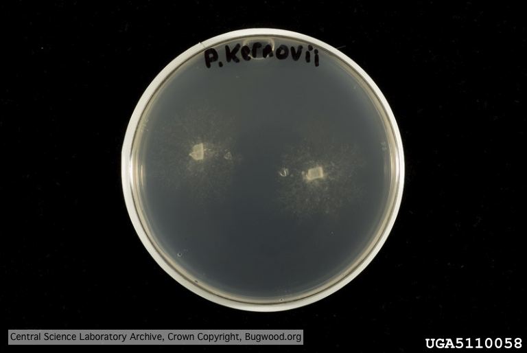 Organism grown on CMA PARP[H]; Plant disease 70, 1038-1043 |
P. cambivora sporangium 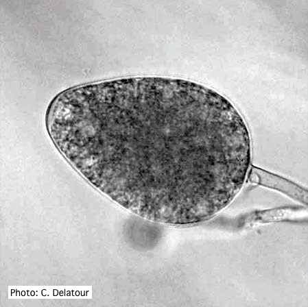 Ovoid non- papillate sporangia |