

Light micrograph of P. agathidicida oospore (Scale bar equals 15 µm)
Photo Gallery
|
P. agathidicia oogonia 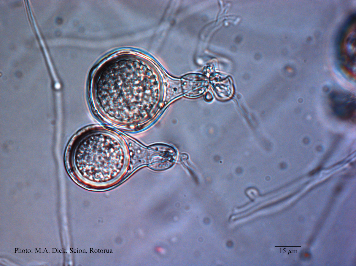 |
P. agathidicia growth on V8  Colony morphology of ex-holotype ICMP 17027 after 10-days incubation at 20°C in the dark |
Mat to control spread of P. agathidicida 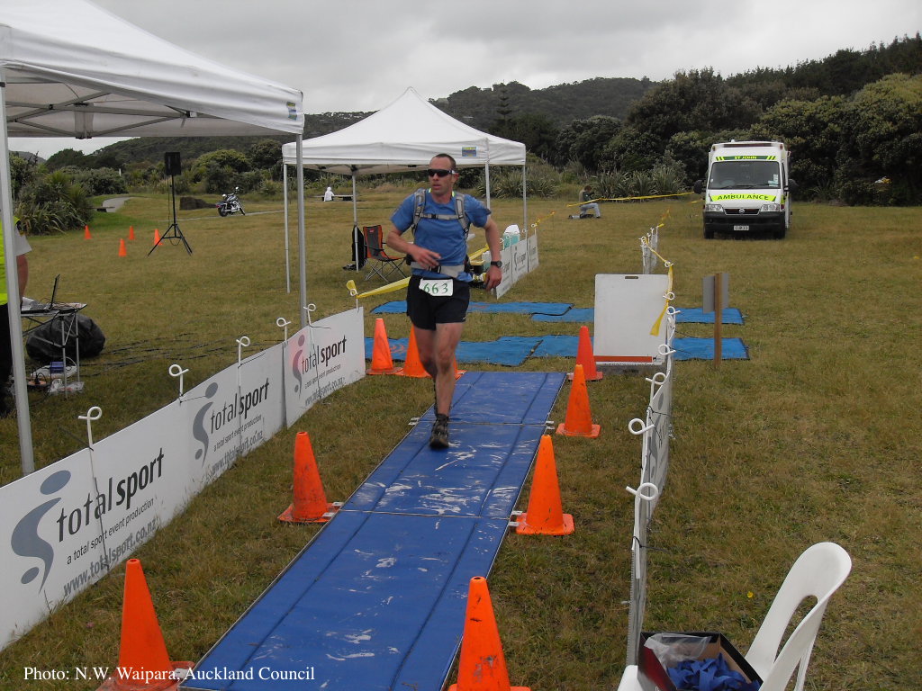 Jogger running over a plastic-reinforced, foam-mat containing a 2% solution of Trigene™ Advance (quaternary ammonium compound) as part of a cross-country event, in the Waitakere Regional Park |
|
Comparative gametangial morphology of Phytophthora Clade 5 species  Comparative gametangial morphology of Phytophthora Clade 5 species, with SEM (top) and light microscopy (bottom). P. heveae has smooth walled oogonia with funnel-shaped, amphigynous antheridia. P. agathidicida has mildly stipulate oogonia with globose amphigynous antheridia. P.cocois has mildly bullate oogonia with reflexed amphigynous antheridia. P. castaneae has coarsely bullate oogonium with rugose protuberances and narrow amphigynous antheridia (Weir et al. 2015). |
P. agathidicia oogonia 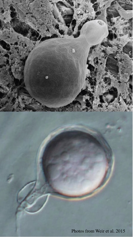 P. agathidicida oogonia with SEM (top) and light microscopy (bottom) |
P. agathidicia disease symptoms on kauri 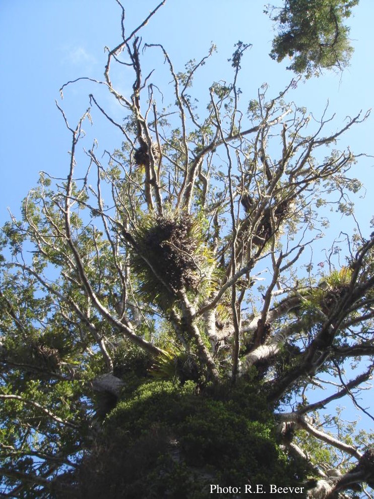 Crown decline of mature kauri, with branchlets with little or no leaves |
|
P. agathidicida oospores in planta  Oospores in the roots of kauri seedlings inoculated with P. agathidicida. The root has been cleared with potassium hydroxide and bleached with peroxide before being stained with trypan blue (scale bar =100 µm). |