

Hyphal swelling photo used with permission from Q-bank
Photo Gallery
|
P. austrocedrae hyphal swellings 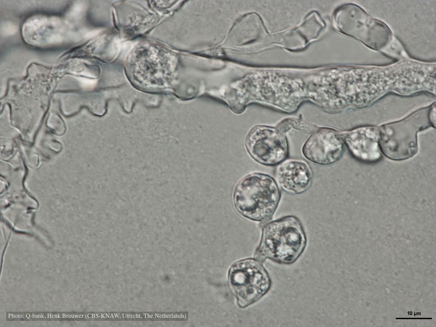 |
P. austrocedrae - hyphal swellings  Morphology of hyphae of Phytophthora austrocedrae, from Greslebin et al. 2007 |
Necrotic lesion in phloem caused by P. austrocedrae  Necrotic lesion in phloem with resin pocket caused by P. austrocedrae |
|
P. austrocedrae colony morphology on Tomato juice agar 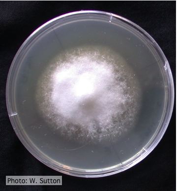 Colony morphology of P. austrocedrae at 16 ºC after 4 weeks on Tomato juice agar |
P. austrocedrae - necrotic lesion in phloem 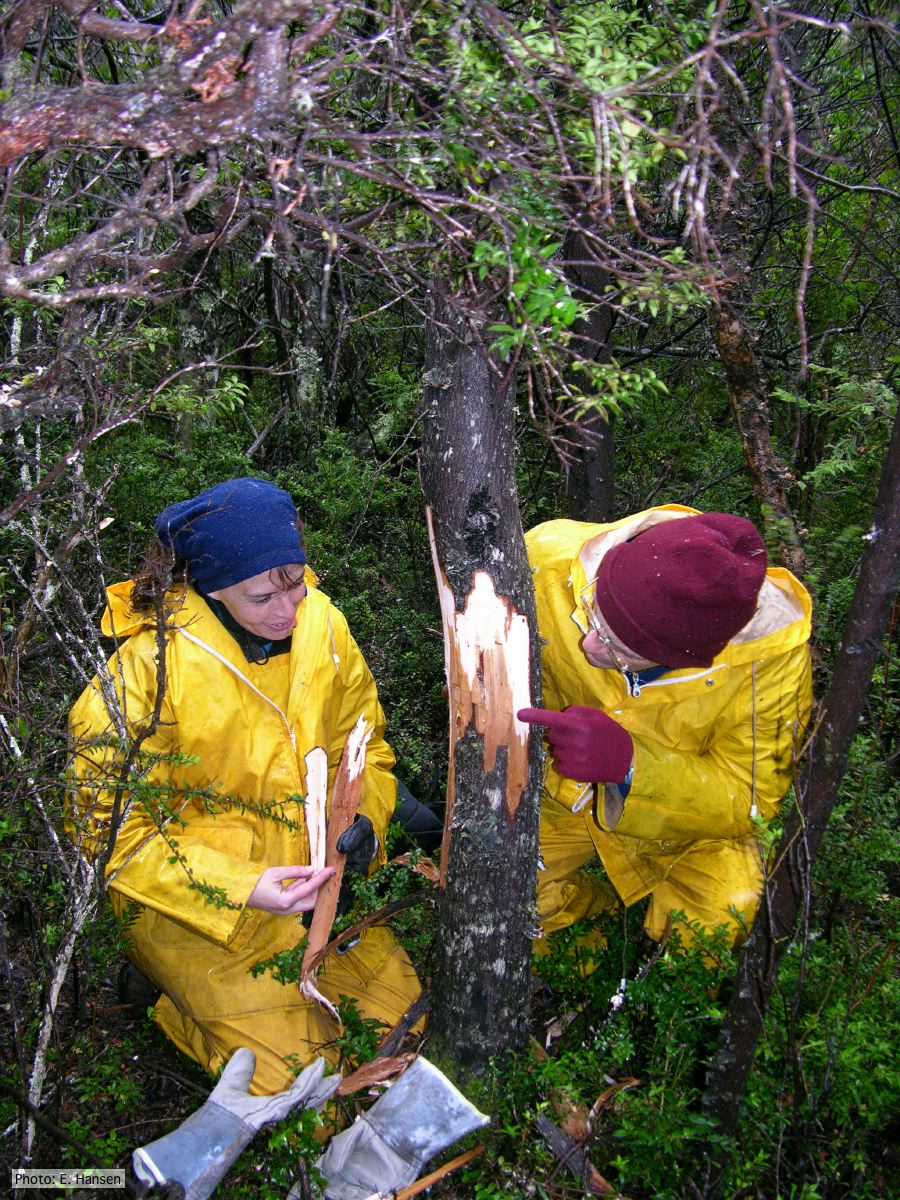 |
P. austrocedrae colony morphology on CMA 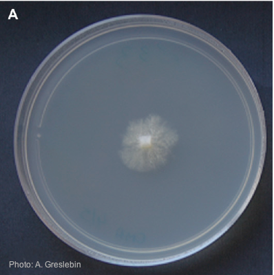 Colony morphology of P. austrocedrae at 16ºC after 4 weeks on CMA |
|
P. austrocedrae oogonium 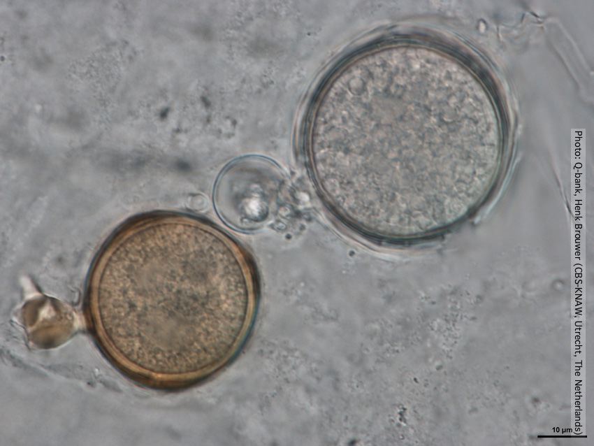 Oogonia with and without brown pigment, photo from Q-bank, used with permission |
P. austrocedrae oogonia drawing  P. austrocedrae. Morphology of oogonia, oospores and antheridia. Bar: 10 mm. Greslebin et al. 2007 |
P. austrocedrae - sporangia 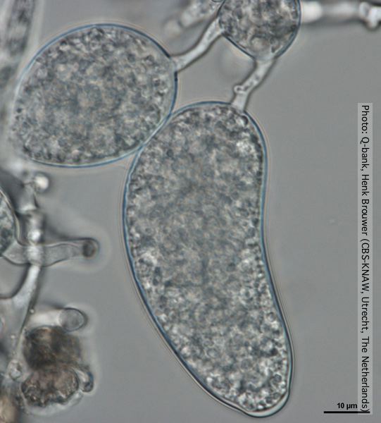 Sporangium with distorted shape, photo from Q-bank, used with permission. |
|
P. austrocedrae hyphal swellings in liquid media drawing  Morphology of hyphae of Phytophthora austrocedrae, from Greslebin et al. 2007 |