

Close-up of margin area of bole lesions under the bark of a bleeding canker
Photo Gallery
|
P. siskiyouensis bleeding canker 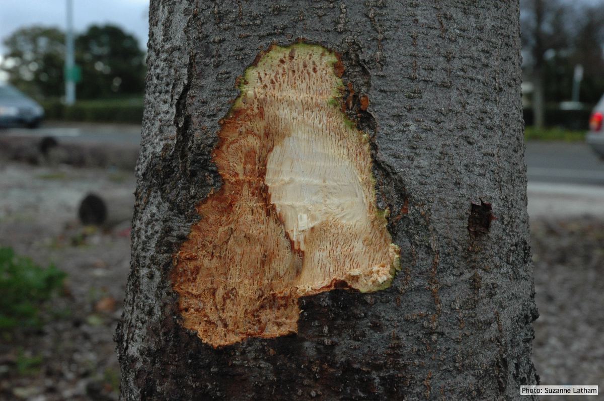 |
P. pluvialis on Pinus radiata 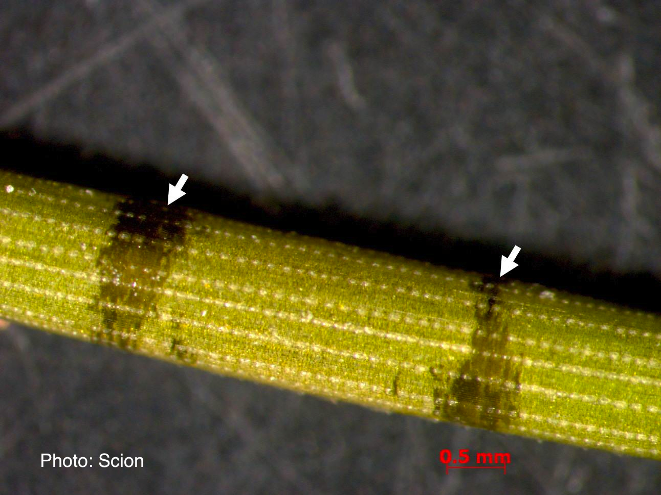 A Pinus radiata needle showing black resinous bands or marks consistent with the presence of red needle cast disease. |
P. chlamydospora colony morphology on carrot agar 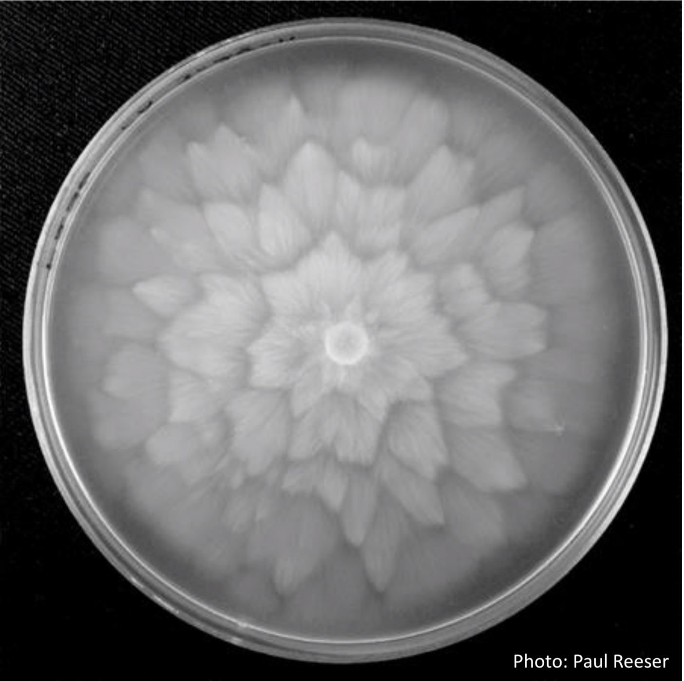 P. chlamydospora colony morphology on carrot agar |
|
P. pluvialis sporangia.  P. pluvialis sporangia on tape peel from infected Douglas-fir needle. |
P. nicotianae chlamydospore 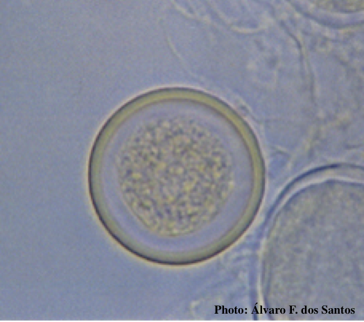 Globose chlamydospore (Fitopatol. bras. 2005) |
P. tentaculata sporangium 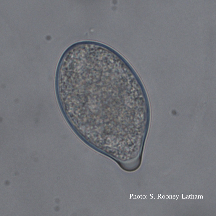 Papillate sporangium of P. tentaculata |
|
P. cactorum bleeding canker 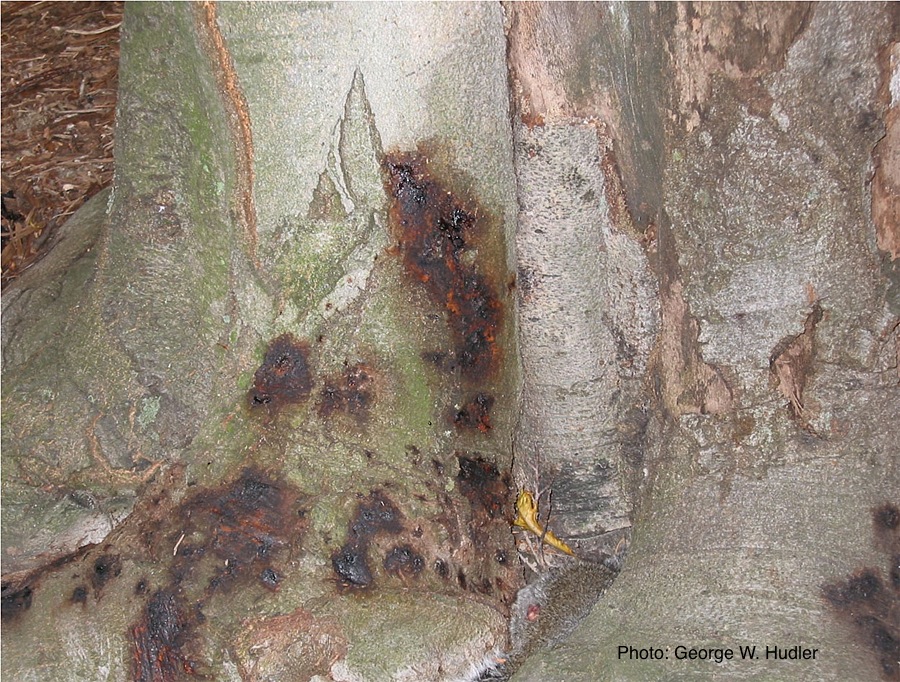 Bleeding canker on European beech (Fagus sylvatica) |
Comparative gametangial morphology of Phytophthora Clade 5 species  Comparative gametangial morphology of Phytophthora Clade 5 species, with SEM (top) and light microscopy (bottom). P. heveae has smooth walled oogonia with funnel-shaped, amphigynous antheridia. P. agathidicida has mildly stipulate oogonia with globose amphigynous antheridia. P.cocois has mildly bullate oogonia with reflexed amphigynous antheridia. P. castaneae has coarsely bullate oogonium with rugose protuberances and narrow amphigynous antheridia (Weir et al. 2015). |
P. tentaculata on V-8 media 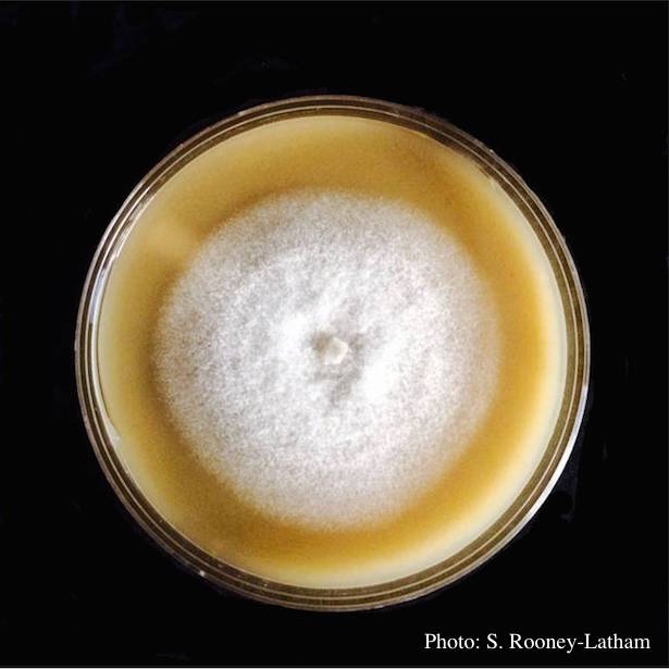 Culture of P. tentaculata on V-8 media |
|
P. pseudotsugae sporangia  Broadly ovoid, papillate sporangia in water |
P. lateralis on Port Orford cedar 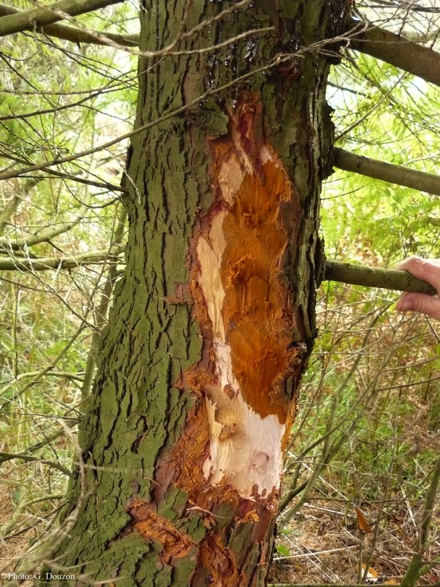 Bole lesion on Chaemacyparis lawsoniana in Lopérec, France |
P. cambivora symptoms 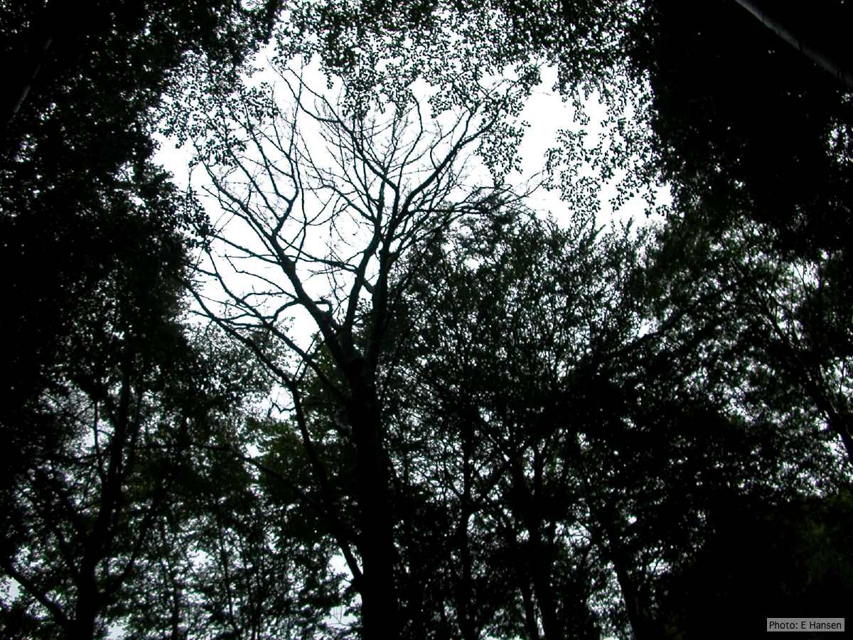 Dead beech in Germany |