

P. ramorum colony morphology on PDA
Photo Gallery
|
P. ramorum colony morphology on PDA 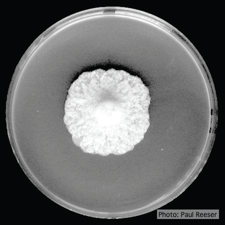 |
P. cactorum colony morphology on PDA 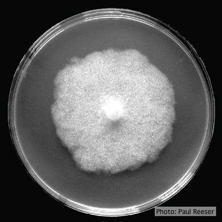 Colony morphology on PDA at 14 days |
P. cambivora sporangium  Ovoid non-papillate sporangia with well-rounded base |
|
P. ramorum zoospores 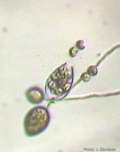 Sporangium of P. ramorum releasing zoospores |
P. cactorum bleeding canker  Bleeding canker on European beech (Fagus sylvatica) |
P. pinifolia coenocytic hyphae 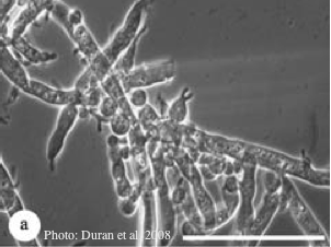 Coenocytic hyphae (from Duran et al. 2008). Scale bar = 20 μm. |
|
P. megasperma oogonium 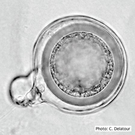 Oogonium with paragynous antheridia applied close to the ogonial stalk. |
P. austrocedrae semipapillate sporangium  P. austrocedrae - semipapillate sporangium with off-center attachment. |
P. kernoviae leaf wilt 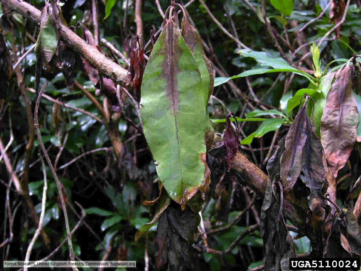 Wilted leaf of infected rhododendron |
|
P. alni symptoms on European Alder  Mature, riparian common alder (A. glutinosa) stand heavily impacted by root and collar rot caused by P. alni |
P. cambivora inactive lesion on chinquapin  Inactive lesion of P. cambivora on chinquapin |
P. palmivora symptoms on fruit  Brown rot on a lemon fruit caused by Phytophthora palmivora. |