

Papillate, non-caducous sporangium with differentiated content; photo used with permission from Q-bank
Photo Gallery
|
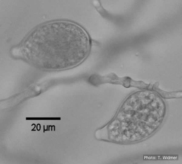
P. katsurae sporangia 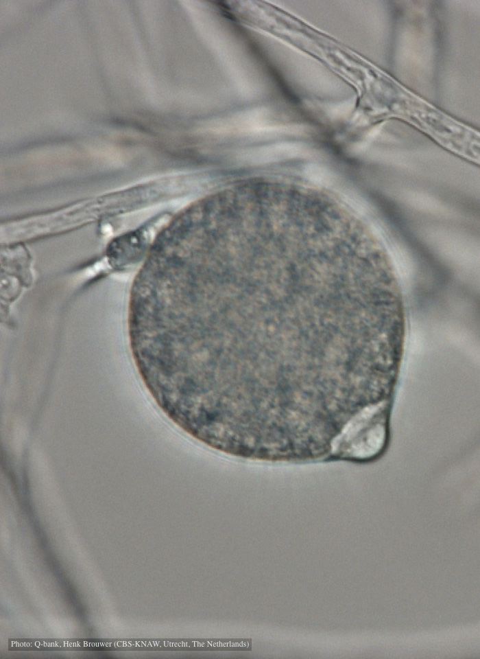 |
P. austrocedrae colony morphology on CMA 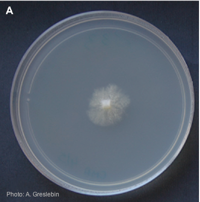 Colony morphology of P. austrocedrae at 16ºC after 4 weeks on CMA |
P. boehmeriae oogonia  Oogonia and oospores with amphigynous antheridia |
|
P. cambivora disease symptoms 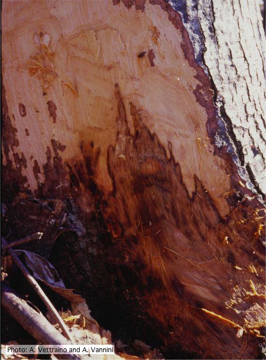 Collar canker rot of Ink disease on sweet chestnut |
P. palmivora sporangia
 P. palmivora caducous papillate sporangia
|
Phytophthora cactorum disease symptoms on English walnut  Dieback of Juglans regia caused by Phytophthora cactorum |
|
Comparative gametangial morphology of Phytophthora Clade 5 species  Comparative gametangial morphology of Phytophthora Clade 5 species, with SEM (top) and light microscopy (bottom). P. heveae has smooth walled oogonia with funnel-shaped, amphigynous antheridia. P. agathidicida has mildly stipulate oogonia with globose amphigynous antheridia. P.cocois has mildly bullate oogonia with reflexed amphigynous antheridia. P. castaneae has coarsely bullate oogonium with rugose protuberances and narrow amphigynous antheridia (Weir et al. 2015). |
P. tentaculata chlamydospore 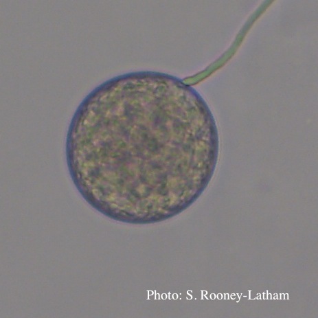 Terminal chlamydospore of P. tentaculata |
P. tentaculata sporangium  Papillate sporangium of P. tentaculata |
|
P. alni subsp alni sporangium 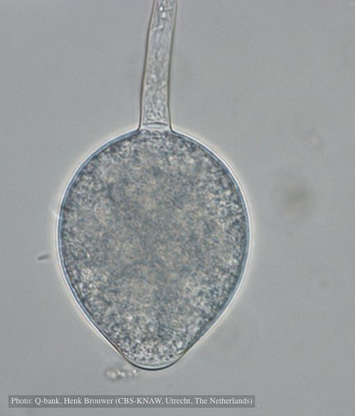 Non-papillate, non caducous sporangium, photo used with permission from Q-bank |
P. cinnamomi hyphal swelling 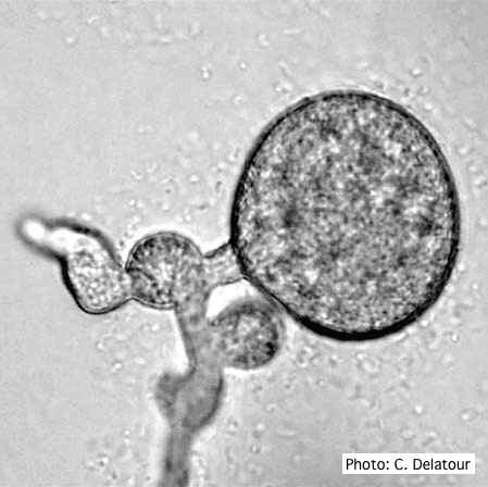 P. cinnamomi hyphal swelling (or thin walled chlamydospores) |
P. agathidicia oogonia 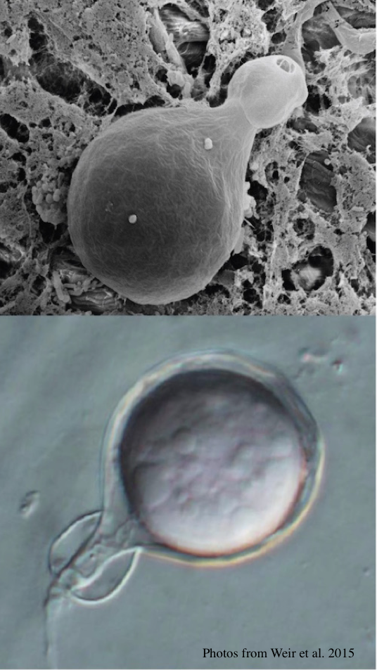 P. agathidicida oogonia with SEM (top) and light microscopy (bottom) |