

Ovoid non-papillate sporangia in water.
Photo Gallery
|
P. cryptogea sporangium  |
P. ramorum zoospores 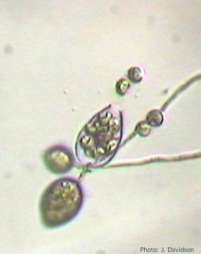 Sporangium of P. ramorum releasing zoospores |
P. siskiyouensis canker on Italian alder  Bleeding canker at the base of a tree and a sprinkler emitter (arrow) adjacent to the trunk |
|
P. pseudotsugae sporangium 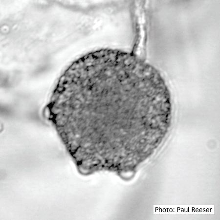 Broadly ovoid, papillate sporangium in water |
P. pseudosyringae sporangia  Ovoid, semipapillate sporangia showing sympodial development of sporangiophore |
P. agathidicida oospores in planta  Oospores in the roots of kauri seedlings inoculated with P. agathidicida. The root has been cleared with potassium hydroxide and bleached with peroxide before being stained with trypan blue (scale bar =100 µm). |
|
P. siskiyouensis bleeding canker  Bole lesions in the tissues under the bark of a bleeding canker: discoloration in the secondary phloem tissue |
P. arenaria sporangia 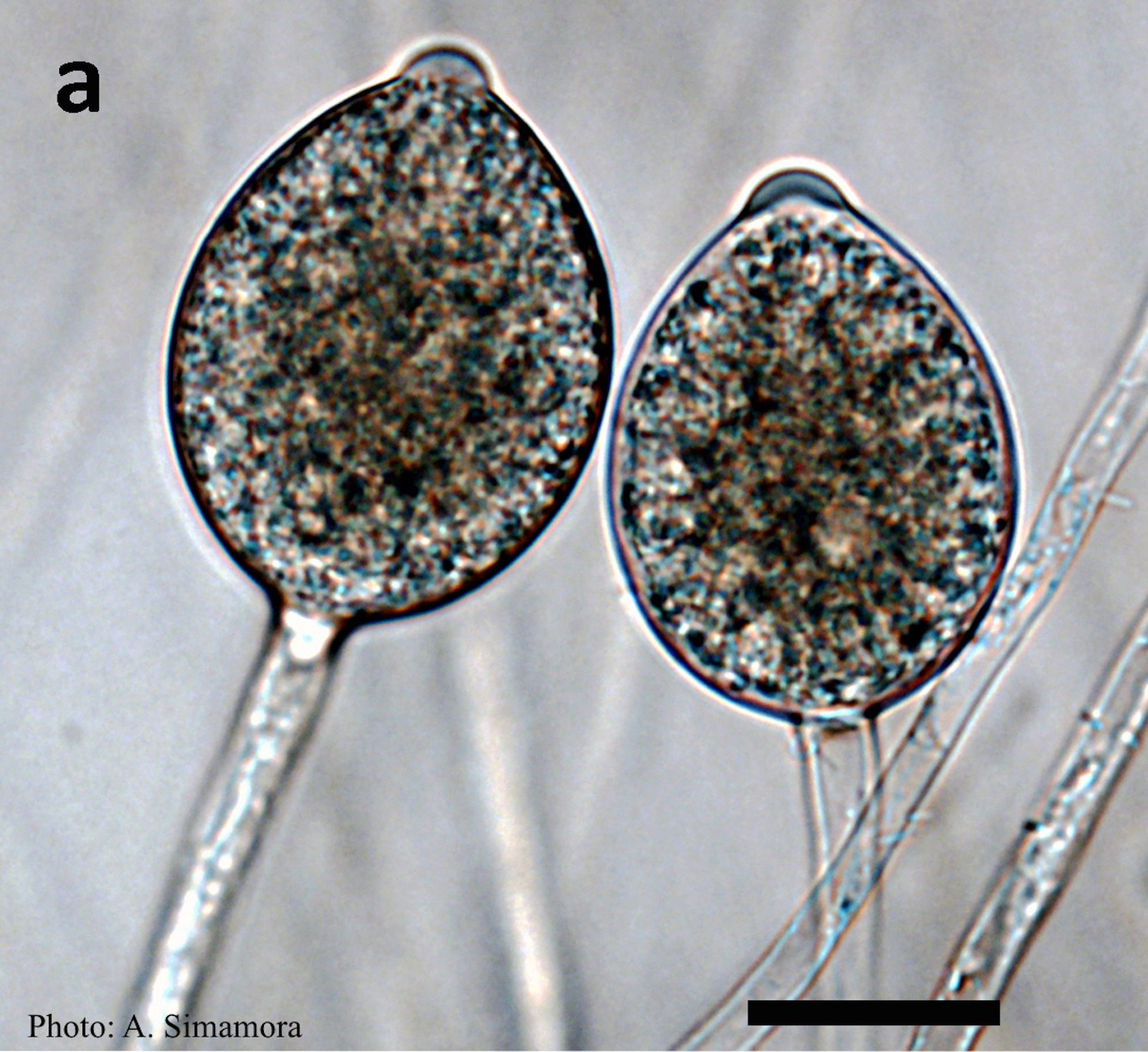 Typical ovoid papillate sporangia of Phytophthora arenaria on V8 agar flooded with soil extract |
Chlamydospore of P. lateralis 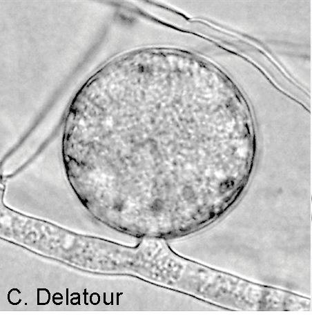 Terminal chlamydospore on a short side stalk |
|
P. pinifolia hyphal growth 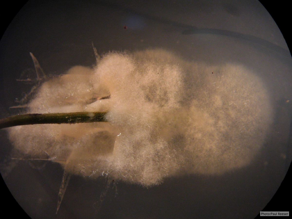 P. pinifolia pathogen growing from infected needle on selective agar |
P. ramorum colony morphology on V8 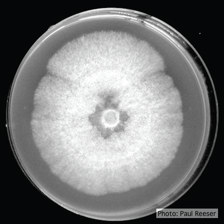 P. ramorum colony morphology on V8 |
P. tentaculata chlamydospore 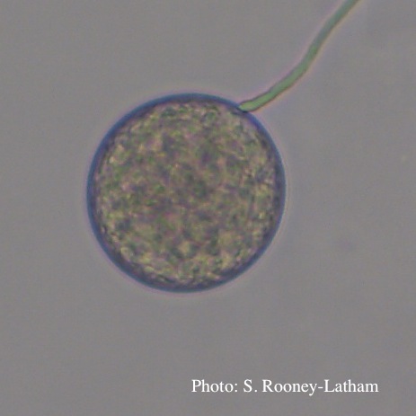 Terminal chlamydospore of P. tentaculata |