

Phytophthora chlamydospora sporangium in water. Bar is 20µm.
Photo Gallery
|
P. chlamydospora sporangium  |
P. austrocedrae - Mal del ciprés, stages of decline 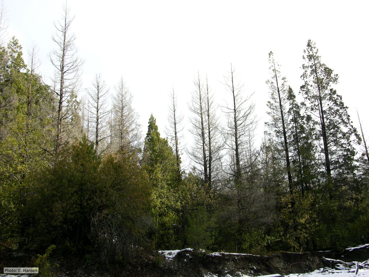 Colony morphology of P. austrocedrae at 16 C after four weeks on PDA |
P. kernoviae sporangium 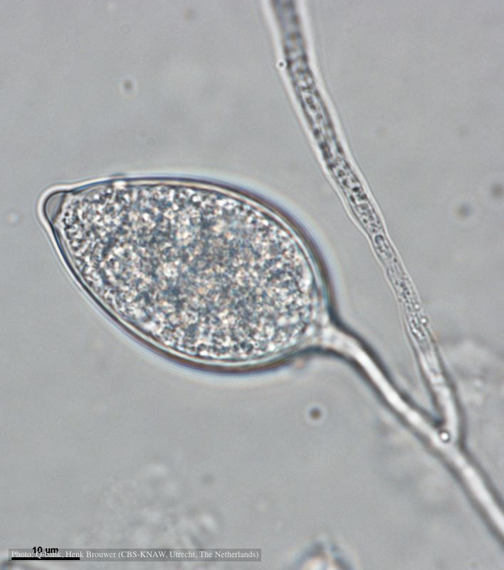 Papillate and caducous sporangium, photo from Q-bank, used with permission |
|
P. cryptogea sporangium  Ovoid non-papillate sporangia in water. |
P. pluvialis hyphal swellings 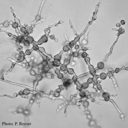 P. pluvialis hyphal swellings on agar |
P. boehmeriae sporangium 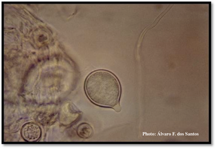 Sporangium showing ovoid and ovoid to spherical shape and papillate condition |
|
P. chlamydospora sporangium 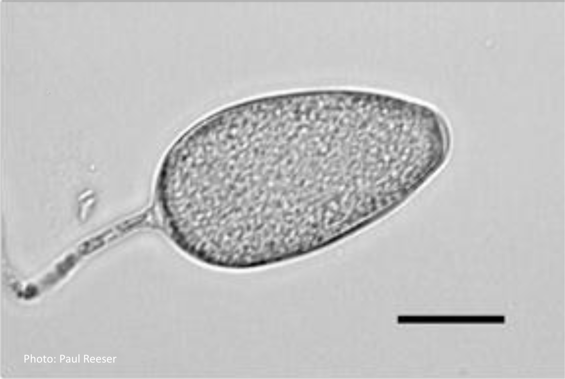 Phytophthora chlamydospora sporangium in water. Bar is 20µm. |
P. pluvialis on Pinus radiata needle  Clusters of sporangia emerge from stomata of an infected radiata pine needle. |
P. austrocedrae hyphal swellings 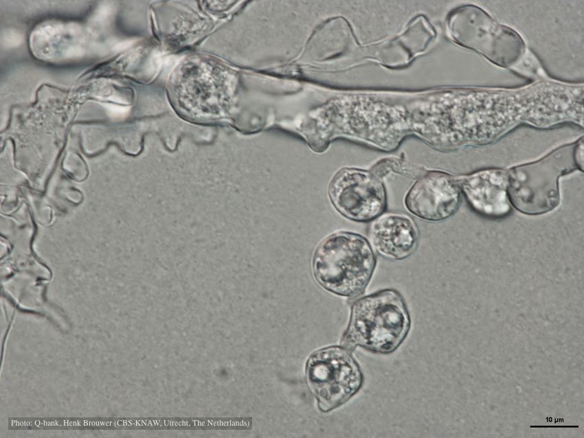 Hyphal swelling photo used with permission from Q-bank |
|
Phytophthora taxon Agathis bole canker  Canker on a Kauri tree, New Zealand |
P. pinifolia hyphal growth 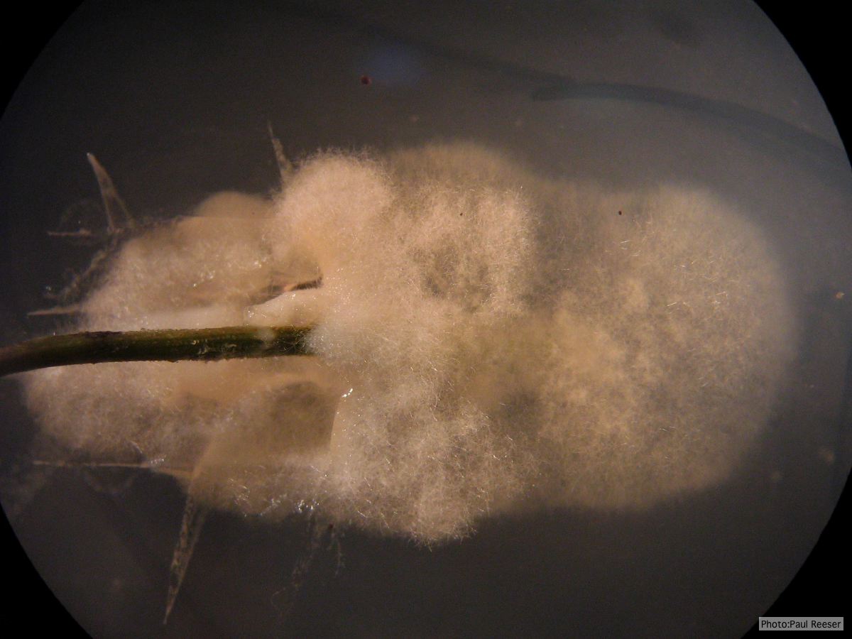 P. pinifolia pathogen growing from infected needle on selective agar |
P. pluvialis hyphal swellings 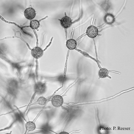 P. pluvialis hyphal swellings in water |