

Papillate sporangium of P. tentaculata with an elongated neck or beak.
Photo Gallery
|
P. tentaculata sporangium 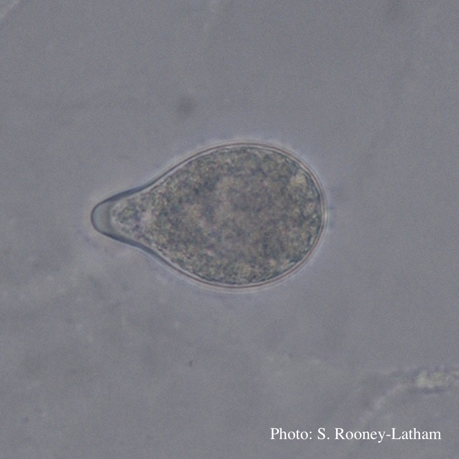 |
Growth morphology on V8 of P. lateralis 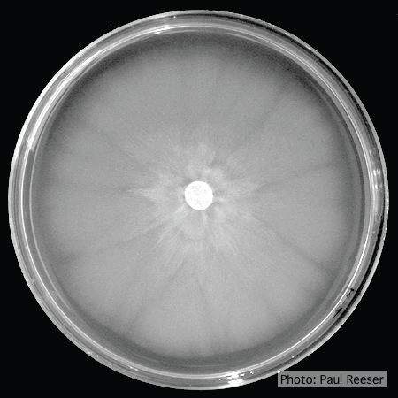 Colony morphology on V8 at 14 days |
P. nicotianae chlamydospore 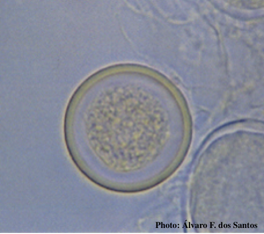 Globose chlamydospore (Fitopatol. bras. 2005) |
|
P. tentaculata disease symptoms on California mugwort  Nursery grown California mugwort plant (Artemisia douglasiana) infected with P. tentaculata and exhibiting severe root and crown rot |
P. alni oogonia subspecies and variants 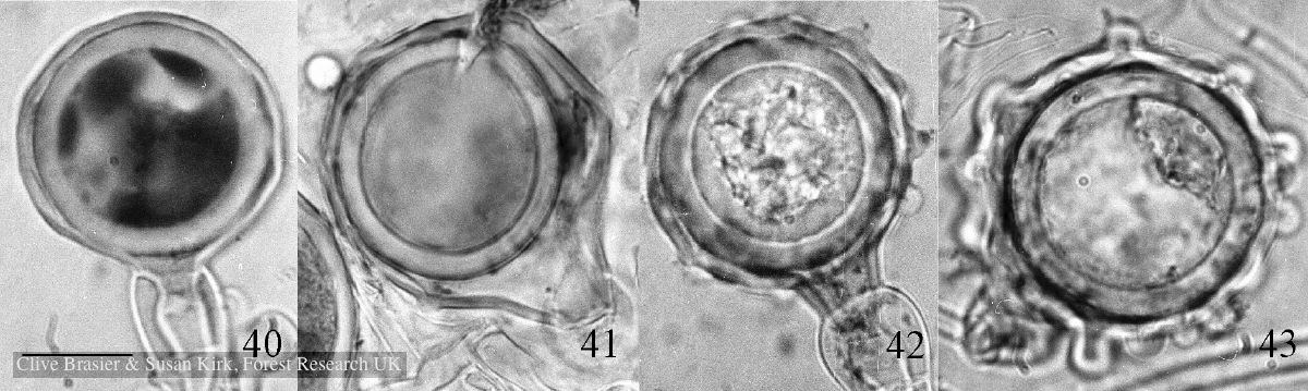 Fig. 40. P. alni subsp. uniformis. Fig. 41. P. alni subsp. multiformis German variant. Fig. 42. P. alni subsp. alni. Fig. 43. |
P. cambivora sporangia 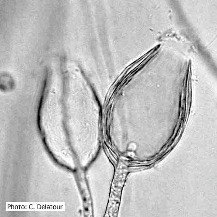 Empty sporangia of P. cambivora showing nested internal proliferation |
|
P. palmivora sporangia, chlamydospores, hyphae 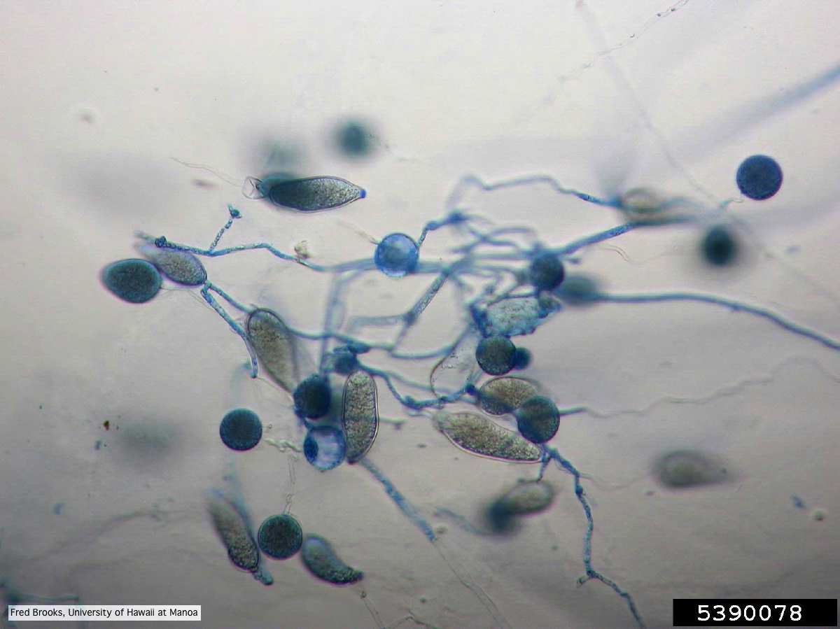 Sporangia (sporangiospores), chlamydospores, and hyphae stained with Cotton Blue |
P. tentaculata chlamydospore 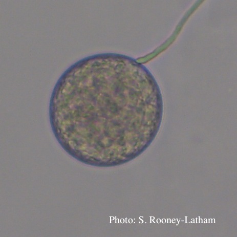 Terminal chlamydospore of P. tentaculata |
Basal canker on Port Orford Cedar stump  |
|
P. lateralis on Port Orford cedar  Localized branch infection of Chaemacyparis lawsoniana in Lopérec, France |
P. alni symptoms on European Alder  Mature, riparian common alder (A. glutinosa) stand heavily impacted by root and collar rot caused by P. alni |
P. katsurae oogonium  Micrograph of warty oogonium |